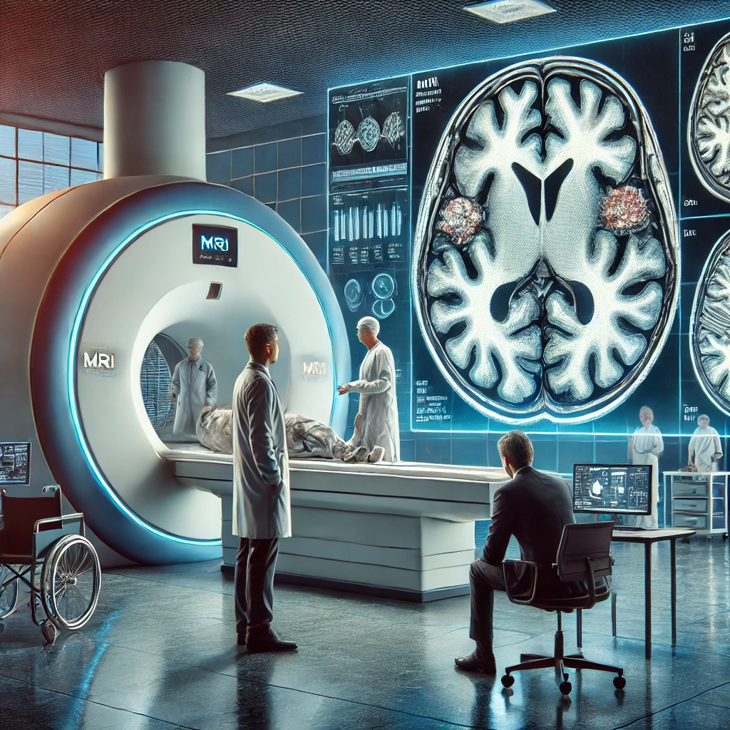
The recent study, A Review of Neuroimaging Findings in Repetitive Brain Trauma, sheds new light on the impacts of repetitive brain trauma, particularly in athletes and military personnel. This comprehensive review, published in Brain Pathology, delves into the advanced imaging techniques used to detect brain anomalies resulting from repeated head impacts. The findings are crucial as they help differentiate between those who recover from such trauma and those who develop chronic traumatic encephalopathy (CTE).
Repetitive brain trauma is a significant concern for professional athletes in contact sports and military personnel exposed to blasts. The study highlights that while all confirmed CTE cases have a history of repetitive head impacts, not all individuals with such trauma develop CTE. This suggests that repetitive impacts might be necessary to trigger the disease, but other factors likely influence its progression.
One of the key takeaways from the study is the potential of magnetic resonance imaging (MRI) to uncover the underlying mechanisms of repetitive brain trauma. The researchers reviewed various neuroimaging findings in athletes and military personnel, revealing how advanced imaging techniques can detect brain changes in both acute and chronic phases of injury.
For instance, diffusion tensor imaging (DTI) has been instrumental in identifying microstructural damage in the brains of professional boxers. Studies have shown that even when clinical MRI scans appear normal, DTI can reveal abnormalities in the corpus callosum and other brain regions. This suggests that repetitive head impacts can cause subtle brain injuries that might not be immediately apparent.
Similarly, a study on high school football players found that repetitive subconcussive head impacts could lead to changes in brain function, even in the absence of a diagnosed concussion. Using functional MRI (fMRI), researchers observed alterations in brain activation patterns, indicating that repeated head impacts might affect brain function over time.
The study also discusses the use of positron emission tomography (PET) to detect tau protein accumulation in the brain, a hallmark of CTE. Recent advancements in PET imaging have allowed researchers to visualize tau deposits in living individuals, providing a potential biomarker for early diagnosis of CTE. This is particularly important as it could lead to interventions that might prevent the progression of the disease.
Interestingly, the study highlights the differences in brain trauma between sports. For example, soccer players, who frequently head the ball, show brain microstructural changes similar to those seen in contact sports athletes. This challenges the notion that only high-impact sports pose a risk for brain injury.
In addition to sports-related injuries, the study also examines the effects of repetitive brain trauma in military personnel. Veterans exposed to multiple blast events during combat have shown brain changes similar to those seen in athletes. For instance, a study on Iraq war veterans found reduced glucose metabolism in certain brain regions, indicating potential long-term effects of blast exposure.
Moreover, recent news highlights ongoing research efforts to prevent brain trauma in athletes. Indiana University researchers are exploring the potential of omega-3 fatty acids to increase brain resiliency to repetitive head impacts. This research could lead to new strategies for protecting athletes and military personnel from brain injuries.
The study emphasizes the need for longitudinal research to understand better the long-term effects of repetitive brain trauma. By following athletes and military personnel over time, researchers can identify early indicators of those at risk for developing neurodegenerative diseases like CTE. This could pave the way for early interventions and personalized treatment plans.
The recent study provides valuable insights into the impacts of repetitive brain trauma and the potential of advanced imaging techniques to detect brain changes. As research continues, it is crucial to develop strategies to protect those at risk and improve the diagnosis and treatment of brain injuries.

